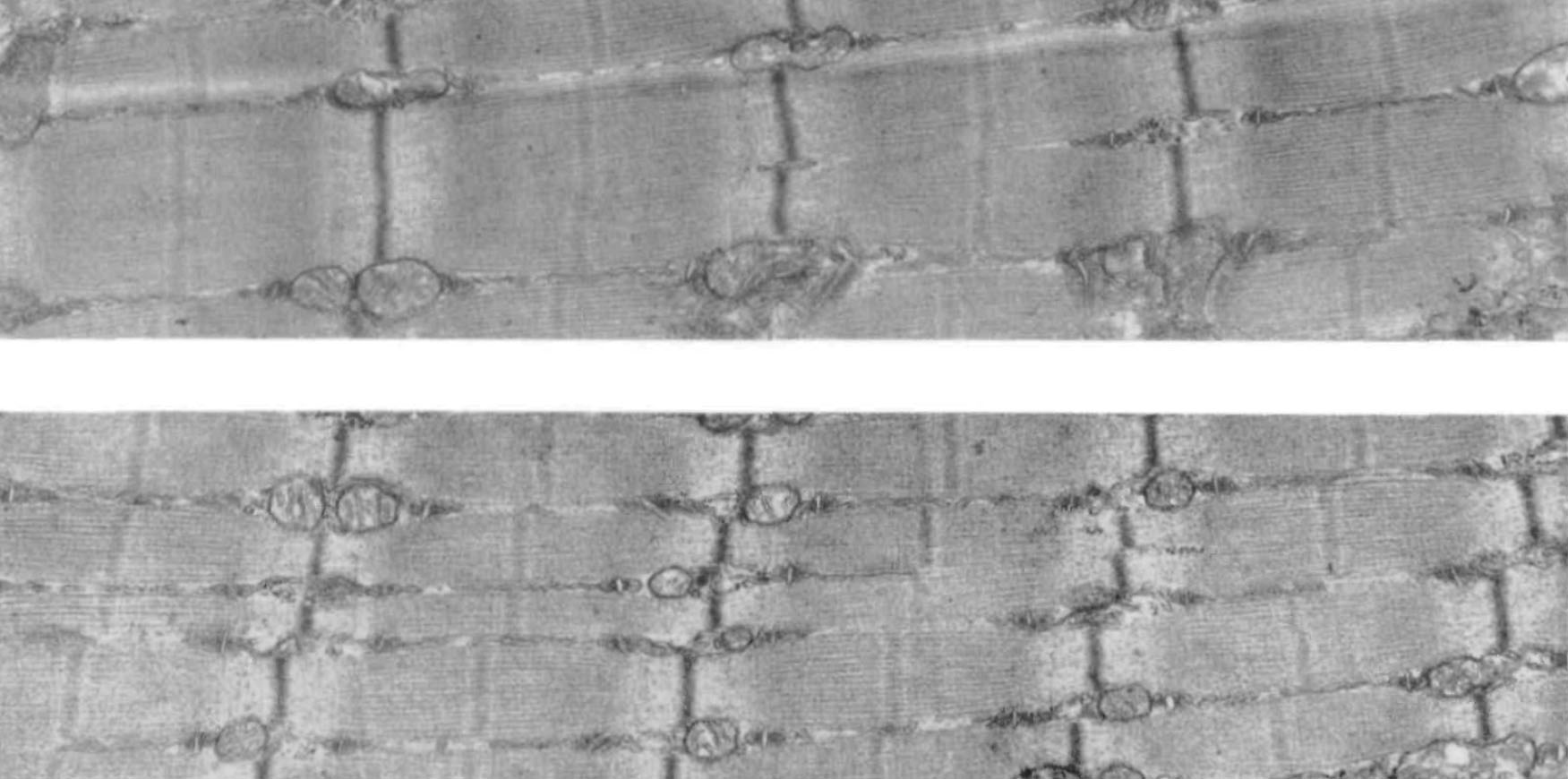
Пролиферация (деление) миофибрилл
При гипертрофии мышц происходит пролиферация (деление) миофибрилл. При помощи электронного микроскопа установлено, что в некоторых средних и крупных мышечных волокнах наблюдается высокая частота продольного расщепления миофибрилл. Измерения в дюймах с помощью электронного микроскопа показали, что миофибриллы, которые расщеплялись, были примерно в два раза больше по сравнению с нерасщепленными миофибриллами и что эти миофибриллы разделялись более или менее по центру.

Голдспинк Г.
Пролиферация миофибрилл во время роста мышечных волокон
С помощью фазового контраста и электронной микроскопии были рассмотрены и измерены миофибриллы в мышечных волокнах различных размеров и разного возраста. Во время послеродового роста двуглавой мышцы плеча мыши количество миофибрилл в некоторых волокнах возрастает от 75 до 1200. Диапазон размеров миофибрилл составляет от 0,4 до 12 мкм. Обнаружено бимодальное распределение размеров миофибрилл в мышцах мышей всех возрастов.
При помощи электронного микроскопа установлено, что в некоторых средних и крупных мышечных волокнах наблюдалась высокая частота продольного расщепления миофибрилл. Измерения в дюймах с помощью электронного микроскопа показали, что миофибриллы, которые расщеплялись, были примерно в два раза большими, по сравнению с нерасщепленными миофибриллами и что эти миофибриллы разделялись более или менее по центру. Возможным объяснением расщепления может быть факт, что тонкие филаменты тянут слегка под углом к центральной оси миофибрилл, из-за расхождения в решетке. Когда миофибрилла достигает определенного размера сила тяги тонких филаментов достаточно сильна, чтобы повредить Z-диск.
Из данных о размерах, форме и числу миофибрилл на разных этапах роста был сделан вывод, что посредством продольно расщепления увеличивается количество миофибрилл во время послеродового роста.
Goldspink, G. The Proliferation of Miofibrils during Muscle Fibre Grows / G. Goldspink // Journal of Cell Science, 1970. – V. 6.– P. 593-603.
Abstract
Myofibrils in muscle fibres of different sizes and different ages were examined and measured using phase-contrast and electron microscopy. During the post-natal growth of the mouse biceps brachii muscle the number of myofibrils in some fibres increases from about 75 to 1200 The range of myofibril size was from 0.4-12 µm.
The distribution of myofibril sizes in muscles of all ages studied was bimodal. A high incidence of longitudinal splitting of myofibrils was observed with the electron microscope in differentiating muscle fibres and in some medium and large muscle fibres. Size measurements with the electron microscope showed that the splitting myofibrils were about twice as large as non-splitting myofibrils and that the myofibrils split more or less down the middle.
A possible explanation for the splitting is that the peripheral I filaments are pulled at an angle slightly oblique to the myofibril axis, because of the discrepancy in the A and I-filament lattice spacings. When the myofibril reaches a certain size the oblique pull of the peripheral I filaments is strong enough to cause the Z disks to rip. From data on the size, shape and number of myofibrils at different stages of growth it was concluded that longitudinal splitting is the means by which the number of myofibrils increases during post-natal growth.
References
- Brandt, P., Lopez, E., Reuben, J. & Grundest, H. (1967). The relationship between myofilament packing density and sarcomere length in frog striated muscle J. Cell Biol. 33, 255-263.
- Carlsen, F. & Knappeis, G. G. (1963) Further investigations of the ultrastructure of the Z disc in skeletal muscle. Acta phystol scand. 59, 213-215
- Fischman, D. A. (1967). An electron microscope study of myofibril formation in embryonic chick skeletal muscle. J. Cell Biol. 32, 557-575.
- Golsdpink, G. (1962). Studies on post-embryonic growth and development of skeletal muscle. 1 Evidence of two phases in which striated muscle fibres are able to exist. Proc. R. It. Acad.62, В id, 135-150.
- Golsdpink, G. (1964). The combined effects of exercise and reduced food intake on skeletal muscle fibres J. cell. comp. Physiol. 63, 200-216.
- Goldspink, G. (1965). Cytological basis of decrease in muscle strength during starvation. Am. J. Physiol. 209, 100—114.
- Goldspink, G. (1968). Sarcomere length during the post-natal growth of mammalian muscle fibres. J. Cell Set. 3, 539-548.
- Goldspink, G. & Rowe, R. W. D., (1968). The growth and development of muscle fibres in normal and dystrophic mice. In Research in Muscular Dystrophy, Proceedings of the 4th Symposium, pp. 116—131. London: Pitman Medical Publishing
- Heidenhain, M. (1913). Uber die Entstehung der quergestreiften Muskelsubstanz bei der Forelle. Arch mikrosk. Anat. Ent zv Mech 83, 427-522.
- Huxley, H. E. (1957). The double array of filaments in cross-striated muscle. J. biophys. biochem. Cytol. 3, 631-648. Reedy, M. K. (1964). Remarks at a discussion on the physical and chemical basis of muscular contraction. Proc. R. Soc. В 160, 458.
- Rowe, R. W. D. & Goldspink, G. (1968). Surgically induced muscle fibre hypertrophy. Anat. Rev 161, 69-76.
- Rowe, R. W. D. & Goldspink, G. (1969). Muscle fibre growth in five different muscles in both sexes of mice. J. Anat. 104, 519-530. Steedman, H F. (i960). Section Cutting in Microscopy. Oxford: Blackwell.
С уважением, А.В. Самсонова